United States Department of Agriculture
AgResearch Magazine


Retinal Scan Technology Identifies
Early TSE Symptoms in Cattle
|
|
Diagnosing and preventing transmissible spongiform encephalopathies (TSEs) is a major priority for U.S. agriculture. Fortunately, ARS scientists have developed and tested a research tool that could be used to screen live cattle for these fatal neurological diseases.
TSEs affect many animals, attacking the central nervous system. The diseases are characterized by misshapen forms of naturally occurring, and usually harmless, proteins known as “prions.” In comparison to their counterparts in healthy animals, prions in infected animals are irregularly folded.
Identifying improperly folded prions is one way to diagnose TSE-infected animals. Another is to examine brain tissue for the riddled, spongy appearance characteristic of TSE-induced brain deterioration. Unfortunately, these tests require that the animal be killed beforehand.
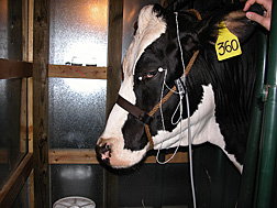
But scientists at the National Animal Disease Center in Ames, Iowa, have determined that retinal scanning technology can detect changes associated with TSE infection in live cattle.
|
|
Led by veterinary pathologist Justin Greenlee, the Ames scientists determined that TSE infection leads to observable changes in cattle retinas. In cattle, as in humans, sight occurs when light enters the eye, and the retina converts it into neuronal signals that the brain interprets as visual images. Within the retina is a type of cell—known as the “bipolar cell”—that plays an important role in this process.
“Bipolar cells take input from the photoreceptors at the back of the eye and hand it off to the ganglion cells that form the optic nerve,” Greenlee explains. “Without the bipolar cells, the message that begins in the photoreceptors never reaches the brain.”
Greenlee’s team included postdoctoral research associate Jodi Smith and ARS technician Leisa Mandell, in collaboration with Iowa State University assistant professor M. Heather West Greenlee. By examining specially stained retinal samples under the microscope, they observed structural changes in bipolar cells in cattle that had been infected with a TSE.
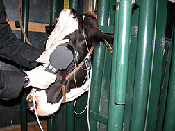
In a later study, the scientists used electroretinography (ERG) to measure retinal response to a flash of light in healthy cattle and in TSE-infected cattle. The scientists observed significant changes in the component of the ERG that measures the response of the bipolar cells, supporting their earlier findings.
In addition, ERG detected these changes about 12.5 months after the cattle had been experimentally infected by intracranial inoculation, about 5.5 months before they generally began to show clinical symptoms.
Now the researchers are investigating how to speed up the testing process and make it more practical for use outside of a laboratory environment.
“Right now, the procedure takes 10 to 20 minutes,” Greenlee says. “An ideal real-world test based on this technology would take just a few seconds.”
Further research is also required to verify that the results can be replicated in animals infected with other TSEs.—By Laura McGinnis, Agricultural Research Service Information Staff.
This research is part of Animal Health, an ARS national program (#103) described on the World Wide Web at www.nps.ars.usda.gov.
Justin Greenlee is with the USDA-ARS National Animal Disease Center, 2300 Dayton Ave., Ames, IA 50010-9684; phone (515) 663-7191, fax (515) 663-7458.
"Retinal Scan Technology Identifies Early TSE Symptoms in Cattle" was published in the August 2009 issue of Agricultural Research magazine.