This page has been archived and is being provided for reference purposes only. The page is no longer being updated, and therefore, links on the page may be invalid.
| Read the magazine story to find out more. |
|
|
|
|
Scientists Rely on High-tech Eyes to Spy on Microscopic World
By Jan SuszkiwAugust 12, 2013
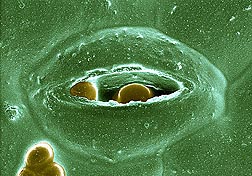
High-resolution images produced by U.S. Department of Agriculture (USDA) scientists at the Electron and Confocal Microscopy Unit (ECMU) in Beltsville, Md., are providing an unprecedented view into the extraordinary world of the unseen.
The images of specimens and samples magnified hundreds of thousands of times their original size have proven invaluable to researchers in describing new pest or pathogen species that pose a threat to U.S. agriculture. On the food safety front, use of the images has helped reveal the mechanisms by which certain bacteria, fungi and parasites infect fresh produce, according to Gary Bauchan, director of ECMU. The unit is part of the Agricultural Research Service (ARS), USDA's chief intramural scientific research agency.
ARS scientists at Beltsville routinely call upon the expertise of Bauchan and the microscopy team to image all manner of specimens and samples.
For example, when a plant pathologist at the ARS National Arboretum's Floral and Nursery Plants Research Unit contacted the team for assistance, the result was a three-dimensional image of how viruses spread from leaf veins to adjacent cells within a leaf. The image, which was generated using fluorescently tagged viruses and a Zeiss 710 confocal laser-scanning microscope (CLSM), illustrated how viruses are transferred within a leaf, and was the cover photograph in the November 2012 issue of the Journal of General Virology.
In addition to the CLSM, the research unit is equipped with a low-temperature scanning electron microscope (LTSEM), a variable-pressure scanning electron microscope, two transmission electron microscopes and a Hirox Digital video microscope. The microscopes are also equipped with digital cameras to speed the delivery of resulting images to researchers.
According to Bauchan, each microscope offers unique capabilities, but requires special handling to prepare specimens and samples prior to imaging. For systematic studies of mites, an LTSEM can be used. This necessitates flash-freezing a mite specimen (while still alive) at minus 321 degrees Fahrenheit and coating it with platinum. This freeze-frames the mite in time, allowing detailed examination of its microscopic features, behavior, and interaction with its immediate surroundings.
Read more about the ECMU and its contributions in the August 2013 issue of Agricultural Research magazine.

