 Image Number D2892-1 |
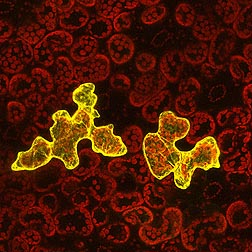
Confocal micrograph of epidermal cells of a plant infected with a virus fluorescently labeled (yellow) to show virus movement from cell to cell within a leaf. Red spheres are chloroplasts from a second cell layer that’s not infected. Image was taken for the U.S. National Arboretum, ARS Floral and Nursery Plants Research Unit.
Photo by ARS Electron and Confocal Microscopy Unit.

